Intestinal-Epithelium
Structure and Development
Section 6.2
(Published August 14th, 2024)
| 🇨🇦 | 🕆 | 🇨🇦 | 🕆 | 🇨🇦 | 🕆 | L🇨🇦 |
6.2.0 Introduction
In the previous section 6.1, we reviewed processes by which enterocytes absorb nutrients from the intestines. In this sub-section, we explore the enterocyte’s birth and life-cycle, as well as that of other closely-related epithelial cells.
Full Disclosure Statement: Please be well aware that I am not a health professional. The information herein is based upon personal life-experiences, research, and knowledge of certain topics which the Lord has graciously guided me through.
Take-note that unique information herein (of which there’s a substantial part) has yet to be evaluated by the health-care industry. As always, heed the advice of your physician and ask for their advice, before varying either diet or lifestyle.
We understand that the human body is comprised of trillions of cells and is quite complex. A little common sense here, reminds us of the fact that we need a certain length of time to change the course of one's health. Not to mention, our daily habits.
Lawrention
6.2.1 The Intestinal-Epithelium Structure
Villus and Villi
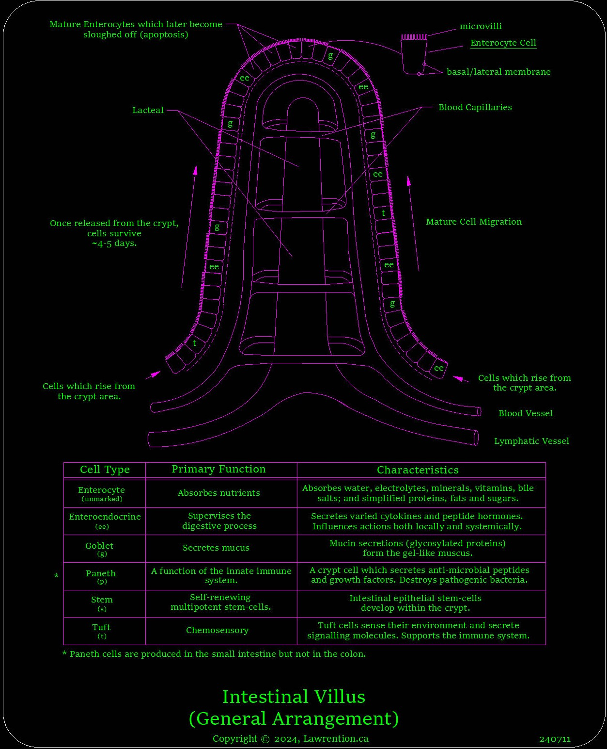
Lining the inner-surface of the small intestine are finger-shaped structures referred to as villi. Villi is their plural term, whereas villus is singular. Each villus is a millimeter long or thereabouts. Take particular note, that although there are absorptive enterocytes within both intestines, there are no villi within the large intestine.
Villi are finger-like protrusions which line the small intestines' lumen. Their primary function is to extract nutrients to nourish our body. Centered inside each villus is a tubular-shaped structure called a lacteal.

The primary role of lacteals, is to absorb fats; fats of which, were previously both ingested and processed by enterocytes. These lacteals connect directly to a network of passages belonging to the lymphatic system.
The lymphatic system is network-like structure that consists of vessels, canals, and organs; of which not only provide areas for transporting dietary fats, but used also for pathogen-defense mechanisms and waste-management.
Each villus has a meshed network of blood capillaries which serves as a bridge between local arteries and veins. These blood capillaries deliver nutrients to cells, and remove waste products from tissues.
Raised clusters of enterocytes which create readily-identifiable profiles - are referred to as brush borders. This term is used due to the fact that the numerous microvilli protruding from the tops of enterocyte cells, look similar to that of hair-brush bristles. These Microvilli sweep away debris from cells, and prepare the way for incoming cellular nutrients.
These epithelial brush-border cells, rise up upon circular-folded regions of the intestinal wall. Approximately, these circular folds are most pronounced within the first two-thirds of the small intestine and end about midway through the ileum.
The brush-border’s shape along with its highly-textured surface that protrudes directly into the (intestinal) lumen, makes it an ideal structure for nutrient absorption.
Commonly, there’s a gel-like substance that coats epithelial cells referred to as glycocalyx. This glycocalyx promotes cell maintenance and cell-to-cell communications; both of which assist the integrity of the brush-border's structure.
Glycocalyx is a composition of interwoven filaments which is advantageous for distancing any quite-large molecular structures away from enterocytes. I believe this glycocalyx serves as basic cell-protection and emerges sometime before the full maturation of its accompanied cell(s).
Now, located above the brush-border and further into the intestinal-lumen; there's a couple layers of mucus (inner and outer layer). This mucus is mainly derived from goblet-cell secretions and supports the gut’s microbiome. Mucus keeps unwanted intruders at bay, while still supporting the inward passage of nutrients.
Sandwiched between the mucus and the intestinal-epithelium - is an aqueous layer referred to as the unstirred water layer. This aqueous layer affects the dynamics of underlying epithelial-cells, and provides them with a certain amount of freedom. Freedom, for moving along the basement membrane, regardless of the fact that these epithelial-cells remain confined under comparatively heavy layers of mucus.
Please take note, that most data which I’ve gathered thus far with regards to the intestinal-epithelium, was typically garnered by those who study rodents. Although some studies may involve humans as well, for obvious reasons - I believe that most of these folks used rodents for examination.
Sample conditions of rodents will vary; and rely upon variables like their species, age, specific section of their intestinal-tract, the rodent's diet; as well as other variables of which I can only imagine. As a result of these various factors, take the information provided herein as a general example; as certain described features likely don’t reflect the true condition of a human's intestine. In particular, the perceived depths of mucus and microbiome growths above the epithelium.
Due to the intestinal-epithelium's unique layered-structure, most gravitational effects towards its cells are effectively negated. Nonetheless, gravity plays a large part in the digestive-process, especially through liquids; but, generally speaking - intestinal epithelium-cells are undeterred by gravity's effects, as one’s body goes through various muscular-contortions and orientations.
Intestinal Crypts
Intestinal-epithelium cells are produced within intestinal-wall glands referred as Crypts of Lieberkühn. These particular crypts are tubular-shaped recessed structures, similar to that of an underground well.
Crypt of Lieberkühn

These Crypts of Lieberkühn are located on comparatively flat regions of intestinal walls, and directly in the midst of protruding villi structures. Ideally speaking, one crypt can support four villi.
Commonly, there are six types of epithelial-cells associated with the Crypts of Lieberkühn, including that of stem cells. These stem-cells are deemed multipotent, since they produce their own niche group of cells.
The cells produced within the Crypt of Lieberkühn are listed below. Their notable characteristics are detailed on the prior Intestinal Villus drawing. From a basic understanding of their primary function, epithelial cells are often categorized as being either absorptive or secretory.
- Enterocyte (absorptive)
- Enteroendocrine (secretory)
- Goblet (secretory)
- Paneth (secretory)
- Stem
- Tuft (secretory)
6.2.2 Intestinal-Epithelium Development
This next bit of text describes certain processes as brush-border cells are being created and released from a Crypt of Lieberkühn. Through dreams and such, and a great deal of research – the description I’m giving will be close…. but not necessarily exact. Therefore, I will state that the following sequence-of-events as cells develop and emerge from the crypt, is “kind of like this”.
Perhaps the best way to understand this series of events, is to backtrack the epithelial-cell developmental process. In so doing, we will be beginning with the best understood processes, and end with the least-understood process; which is likely that of stem-cell activity.
There are four types of brush-border cells which populate villi. These are the enterocyte cells which ingest nutrients; enteroendocrine cells which supervise and regulate the digestive process; goblet cells which secrete mucus and support the mucus layers above the brush-border; and the tuft cells which sense their environment and release signaling molecules to the immune system. These four types of cells migrate up their respective villus, and beneath the cover of mucus (both inner and outer mucal layers).
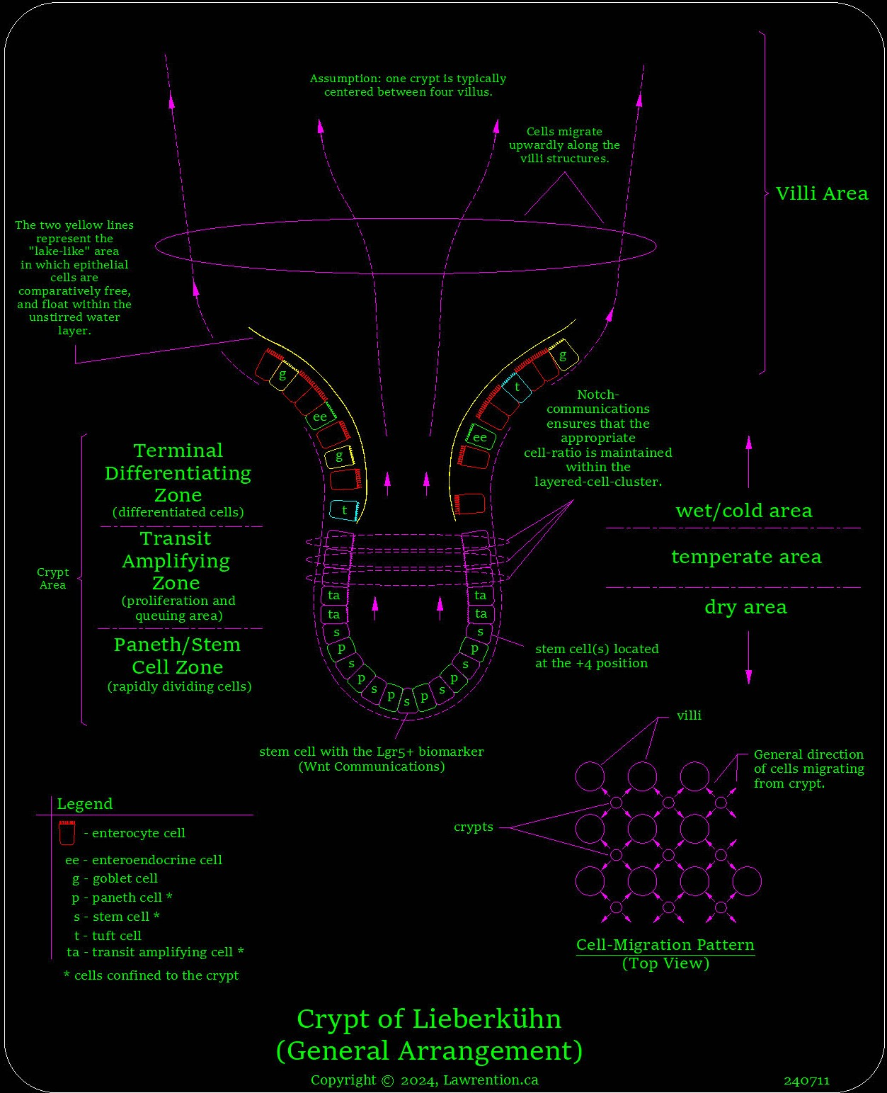
It seems that two-dimensional drawings depicting the migration of epithelium-cells from the crypt to the brush-border are naturally limiting; as its difficult to grasp the true dynamics of both cell proliferation and migration. Please refer now to the following drawing.

In the bottom right corner of the drawing, there’s a cell-migration pattern showing how epithelial cells move from the crypt to associated villi. Here we see that each Crypt of Lieberkühn supplies cells to four different villi structures. Now, likely not all villi/crypt arrangements are this symmetrical; but, I believe that this cell-migration pattern is typical.
Brush-Border Communications
The epithelial cells which migrate up each villus’ profile in a coordinated and progressive fashion, later evolve into the brush border. While doing so, communication to neighboring cells occur through close contact, and most-likely via the common Wnt and/or Notch-signaling pathways. Both Wnt and Notch-signalling pathways are common means for cell-to-cell communications.
The enteroendocrine cells which regulate and supervise much of the intestinal-epithelium processes - communicate not only locally (to neighboring cells via cytokines); but also systemically, by releasing hormones into the blood-stream. In addition, there are studies being done to determine how these cells communicate to the enteric nervous system (ENS) through their basement membrane.
From what I understand, enteroendocrine cells have varying subtypes as well. Enteroendocrine subtypes accommodate for differing hormone secretions which can have varying effects upon the digestive process.
I believe that in many cases, a specific enteroendocrine subtype is a direct result of the intestinal-region in which it emerges from the crypt. In addition, it's been determined that differing enteroendocrine hormone-secretions can occur merely from changing the type of foods in which they encounter.
So, as it seems - enteroendocrine cells have the ability to change their secreted type of hormone, merely by changing the nutritional content within the intestinal-lumen. As a result of this feature, these enteroendocrine cells become an important part of the central-nervous-system / enteric-nervous-system bi-directional communication link. This bi-directional link is referred to as the gut-brain axis (GBA).
Studying this gut-brain axis is complex, and minimally involves topics such as those listed below.
- Gastrointestinal homeostasis.
- The hunger/satiety response.
- Emotional effects related to the gut's microbiota.
As unusual as this seems, I also understand that enteroendocrine cells within the gut provide similar functionality as the mouth's taste-buds. So, there's much here to "chew on" so to speak, when it comes to the gut-brain axis.
Cell-Migration from Crypt to Villi
Between the brush-border and the (two) mucal layers lining the intestinal-lumen, there’s an unstirred water layer. This unstirred water layer provides a means for epithelial-cells to have a certain amount of freedom while they migrate along the basement membrane.
I believe that the (two) mucal layers above the unstirred water layer are contiguous like a blanket, and spread outwardly from the crypt's mouth towards nearby villi. These gel-like layers are supported by mucus-secreting goblet cells that periodically emerge from the crypt. Essentially, these mucal layers trap the fluid within the unstirred-water-layer.
The unstirred-water-layer of the basin area (which is the comparatively flat area between crypt and villi) is normally highly saturated with water. Due to the minuscule cell-size of those being released from the crypt, comparatively speaking - the body of water surrounding the crypt looks like a lake.
The water within this basin-area lake serves as a transition and buffering zone between the crypt and nearby villi; so, a perfectly-timed emergence of cells from the crypt is really not necessary. Potential gaps in this basin area which often become void of epithelial cells, get immediately filled with water.
As a result of this virtual lake between the crypt and corresponding villi - the timing of cells released from the crypt is somewhat sporadic. Further, the crypt cell-production-rate is not tightly coordinated with specific villi, as there’s no need to do so as might be suggested by others.
As a host of epithelial cells remain in one of these basin-area lakes, they float independently of one another like small boats. While doing so, there’s no need for cell-to-cell communications and likely not necessary until they begin to climb a villus structure.
Again, this basin-area-lake is a trapped layer of water located above the basement membrane and beneath the overhead mucus layer(s). This body of water serves as a buffering area, for a fast migration of epithelial cells between the crypt and the base of villi. Reasoning this through, I can’t see any reason for soon-to-be brush-border-cells to quickly migrate across this basin-area lake, and later travel "uphill" towards villi structures, other than by using various rates of electron flow.
Here, it's quite likely that these epithelial cells' motility is due to an electrical-gradient just beneath the mucal layer(s). Commonly understood, is the fact that mucus within the intestines has a blanketing effect and has an overall negative electrical charge.
Cell Emergence from The Crypt’s Mouth
The crypt’s job is to prepare lineage in order to ensure there’s an appropriate ratio of varying epithelial cell-types. The area in which these cells emerge is referred to as the Terminal Differentiating Zone.
Each of these crypts can be considered as being equally divided into three distinct zones: the Terminal Differentiating Zone, the Transit Amplifying Zone, and the Stem/Paneth Cell Zone. Each of these zones are shown on the following drawing.
As a guess to the ratio of cell-types that emerge from the crypt - perhaps for every ten enterocytes; there could be two goblet cells, one enteroendocrine cell, and one tuft cell.

Within the central area of the crypt, we have what’s referred as the Transit Amplifying Zone. Here, the mouth of the crypt narrows, and forces proliferating cells into a ring-like cluster.
As cells move upwardly towards this narrowing mouth of the crypt, they are naturally squeezed together. As a consequence, they become interconnected through proteins referred as Notch signaling pathways. Notch-signaling pathways accommodate cell-to-cell communications, and arise simply due to proximal cell-cell compression, and the subsequent interlocking of transmembrane-proteins.
Within this transit amplifying zone, I believe that height-wise there’s at least three different cell layers which get squeezed together in order to form notch-signaling pathways. As a result, a cluster of cells within the crypt’s mouth communicate together as a single unit. As we progress through this narrative, I will merely refer to this cluster of cells in the mouth of the crypt as the “layered cell cluster”.
Now, the base of each Crypt of Lieberkühn is comparatively dry. Water doesn’t travel into the crypt itself from above, as there’s a seal of sorts near the crypt’s opening. I'm assuming that the mucus which wraps around the crypt's mouth, and is immediately above the unstirred-water-layer, works as a semi-permeable seal.
An epithelial cell cannot be released from the layered-cell-cluster until enough water reaches its upper edge, and thereby lifts and floats a cell away. This action may be analogous to that of a rising-tide that approaches a boat upon the beach.
As this soon-to-be-free cell begins to float, its notch-communication link (or links) which were previously attached to an adjacent cell (or cells) become severed.... and the cell floats freely away. Shortly thereafter, the remaining layered-cell-cluster checks for the appropriate cell-type-ratio, and makes the proper adjustment(s).
On the right side of the Crypt of Lieberkühn drawing, I’ve identified three distinct crypt areas based upon its ambient conditions. The moisture content within the temperate area serves as an important factor in regulating the ongoing process of cell-differentiation. Perhaps the final determination of a specific cell-type doesn't occur until the cell reaches the upper-most layer of the cluster.
Terminal Differentiating Zone (upper-third of crypt)
From my current impression, as cells emerge from the layered-cell-cluster, their cell-type has already been determined. Once a cell is released from the mouth of the crypt and enters the (basin-area) "lake", they become saturated with water and fully mature. Here, there's likely more than ample time allowed, so an epithelial-cell can mature independently before climbing a villus structure.
This basin-area-lake, is likely where goblet cells mature and begin to produce mucus in order to support the mucal layer(s) immediately above the unstirred-water-layer.
Transit Amplifying Zone (center-third of crypt)
The transit amplifying zone is the central third area within the Crypt of Lieberkühn. Here, the transit amplifying cells (TA cells) which rise from the crypt, later mature into one of four types of cells (enterocytes, enteroendocrine cells, goblet cells, and tuft cells). Due to this niche group of cells which directly evolve from TA cells, these TA cells may be classified as multipotent stem cells.
As I stated within the earlier section on cerebral spinal fluid (Section 5.1.2), one way to envision how stem-cells function, is by borrowing a term from the science of physics, which is superposition.
Here, each stem cell experiences what might be considered as an identity crisis before differentiation.
The transit amplifying zone as a whole, is a queuing-up area where cells begin to differentiate. In order for a TA cell to migrate upwardly along the wall of the crypt, it has to first pass a “quality test” of sorts. This ensures that its multipotent character is perfectly capable of differentiating into its expected progeny.
Stem/Paneth-Cell Zone (bottom-third of crypt)
There are two types of cells in the base of the crypt which supports intestinal epithelial-cells. These are the paneth cells and crypt-niche stem-cells. Within this area a competition of sorts ensues, whereas a cell has to reach a certain level of maturity before advancing upwardly and becoming a TA cell.
I believe that there can be up to a dozen paneth cells at any one-time within a Crypt of Lieberkühn. For some reason, paneth cells proliferate with the crypts of the small intestine; but are not expressed within the colon.
Paneth cells secrete peptides and proteins which work to defend against bacteria and other potentially harmful microorganisms. Paneth cells can also function as macrophages. Hence, the assemblage of their varied skill-set - garners them the power to support both the gut's immune system and the microbiome.
We will now discuss the perplexing topic of stem cells. To date, I haven't been able to string together the whole series of events which lead to the emergence of self-renewing stem cells. Nonetheless, I do know a thing or two about mechanisms which ultimately govern their metabolism and lineage's rate-of-production. In other words, a cell's "stemness".
Stemness characterizes an undifferentiated cell type as having the ability of creating a different type of cell, and one that's distinguished from the prior. Studies show that a stem cell will lose its stemness once removed from the crypt. Curiously, its been found that as a differentiated cell is returned to the crypt... its stemness can miraculously return.
In regards to current knowledge for that of stem-cell activity and their lineage rate-of-production - I believe that the mysterious topic of morphogens is basically at the tip of the spear.
These Morphogens are signaling-molecules which can influence their environment (molecules, cells, and the like) merely due to their concentration. From what I understand, it’s a morphogen-concentration-gradient that determines both the threshold and rate of stem-cell activity.
It appears that upon reaching a certain morphogen concentration, they can communicate to each other through some (as of yet) undermined means. As a result of this poorly-understood deep methodology of matter - morphogenic experiments can be difficult to repeat.
I gather that during these morphogen experiments variations often creep in, regardless of the fact that biological-wise and molecular-wise - one experiment seems identical to another.
Further, although we might recognize certain biological and molecular structures, such as a unique protein, molecule, or perhaps even a chemical element; we cannot adequately study something that's more fundamental in nature. As a result, certain investigations attempting to unveil these type of mysteries, might be considered as a type of shell game.


